Wissenschaft

Wissenschaft & Forschung

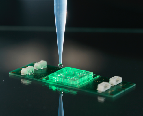
Nicht wachstumsbasierte Methoden zur phänotypischen Untersuchung von Mikroorganismen bieten einen schnellen und markierungsfreien direkten Zugang zur Identitätsbestimmung, zu lebenswichtigen Stoffwechselfunktionen und zur Untersuchung mikrobieller Wechselwirkungen mit Substanzen. Akademische Anwendungen des softwaregesteuerten vollautomatischen modularen Gerätekonzepts GramRay.

Die Raman-Spektroskopie unterscheidet sich von anderen derzeit angewandten Techniken durch ihre einfache Anwendung zu niedrigen Kosten, ihre hohe Analysegeschwindigkeit und ihren breiten Informationsgehalt sowohl über die chemische Zusammensetzung als auch über die Struktur von Biomolekülen innerhalb der Mikroorganismen.

Leichte Veränderung der chemischen Zusammensetzung von Mikroorganismen kann mit Hilfe der Raman-Spektroskopie überwacht und zur Differenzierung von Gattungen, Arten oder sogar Stämmen verwendet werden. Der Nachweis von Krankheitserregern ist aus komplexen Matrices wie Boden, Nahrung und Körperflüssigkeiten möglich. Darüber hinaus werden spektroskopische Untersuchungen von Wirt-Pathogen-Interaktionen sowie die Wirkung von Antibiotika auf Bakterien behandelt.

Akademische Anwendungen nutzen die höchste Empfindlichkeit bei der Einzelzell-Identifizierung für die Untersuchung von Zoonosen und den Nachweis von Krankheitserregern aus Getränken und Lebensmitteln. Vibrionen wie z.B. Cholera sind lebende, schwer kultivierbare Keime (VBNC), die mit GramRay ohne vorherigen Kultivierungsschritt zuverlässig nachgewiesen werden können. Ihr Stoffwechsel wird mit stabilen Isotopen Raman (SIRM) untersucht. Bei der Aufklärung von Mikrobiomen liefert GramRay einen direkten Zugang zu Populationen, da Wachstums- und Inkubationsschritte entfallen. Raman zeigt auch nicht PCR Primer spezifische Lücken, wie z.B. das breit eingesetzte Next Generation Sequencing Verfahren zur Aufklärung des Mikrobioms.

Mit dem GramRay-System kann die Lebensfähigkeit von Bakterien direkt untersucht werden. Die Wechselwirkung mit Substanzen oder Wirts-Pathogen Interaktionen sind sofort sichtbar. So kann die Eignung potenzieller Antibiotika für spezifische Bakterien innerhalb von nur 1 Stunde bestimmt werden. Durch die Visualisierung der Interaktion eines Antibiotikums mit der Bakterienmembran werden die Mechanismen der Antibiotikaresistenz aufgeklärt.

Merkmale und Vorteile

  • Probenvolumen: 0,1 – 500 ml
  • Arbeitszeit für den Laboranten: 10-30 min
  • Empfindlichkeit: 10 Mikroben in Abhängigkeit von der Matrix und Seperationsverfahren
  • Zeit zum Ergebnis: 90 min
  • Zählung der Gesamtkeimzahl: 15 Minuten
  • Zeit für die Gattungs-/Arten-Klassifikation: 15 s pro Mikrobe
  • Integrierte erprobte Software zur Modellbildung  spezifischer chemometrische und maschinelle Lernmethoden:
    • lineare Diskriminanzanalyse (LDA)
    • Random Forest (RF)
    • k-nächste Nachbarn (kNN)
    • Support-Vektor-Maschine (SVM)
    • partielle kleinste Quadrate der Diskriminanzanalyse (PLS-DA)
    • unüberwachte hierarchische Ward-Cluster-Analyse (HCA)
    • lineare Regression; partielle kleinste Quadrate der Regression (PLSR)..
  • Integrierte Validierungsalgorithmen:
    • leave-one-batch-out Kreuzvalidierung (LOBOCV)
    • 10-fache Kreuzvalidierung
  • Flexible Probenmenge
  • Effizientes arbeiten und schnelle Herstellung viele Proben
  • Automatisierung erhöht die Reproduzierbarkeit der Forschungsergebnisse
  • Höchste Empfindlichkeit und Genauigkeit: jede lebende Zelle wird gezählt:
    • Differenzierung zwischen: Tot/Lebendig/VBNC
  • Hoher Probendurchsatz und Spektren Generierung zur Modellbildung
  • Benutzerhierarchie und Datenintegrität ermöglicht die Rückführbarkeit von Versuchsergebnissen

Auszug aus der Veröffentlichungsliste der GramRay technologie:

  • S. Stöckel, S. Meisel, R. Böhme, M. Elschner, P. Rösch and J. Popp, ” Effect of supplementary manganese on the sporulation of Bacillus endospores analysed by Raman spectroscopy”, J. Raman Spectrosc. 2009, 40, 1469-1477.
  • S. Meisel, S. Stöckel, M. Elschner, P. Rösch and J. Popp, “Assessment of two isolation techniques for bacteria in milk towards their compatibility to Raman spectroscopy”, Analyst 2011, 136, 4997-5005.
  • S. Stöckel, S. Meisel, M. Elschner, P. Rösch and J. Popp, “Raman Spectroscopic Detection of Anthrax Endospores in Powder Samples”, Angew. Chem. Int. Ed. 2012, 51, 5339-5342.
  • S. Meisel, S. Stöckel, M. Elschner, F. Melzer, P. Rösch and J. Popp, “Raman spectroscopy as a potential tool for the detection of Brucella spp. in milk”, Appl. Environ. Microbiol. 2012, 78, 5575-5583.
  • S. Stöckel, S. Meisel, M. Elschner, P. Rösch and J. Popp, “Identification of Bacillus anthracis via Raman spectroscopy and chemometric approaches”, Anal. Chem., 2012, 84, 9873−9880.
  • U. Münchberg, L. Wagner, E. T. Spielberg, K. Voigt, P. Rösch and J. Popp, “Spatially resolved investigation of the oil composition in single intact hyphae of Mortierella spp. with Raman micro-spectroscopy”, Biochim. Biophys. Acta Mol. Cell Biol. Lipids, 2013, 1831, 341-349.
  • S. Pahlow, S. Kloß, K. Kirsch, U. Hübner, P. Rösch, K. Weber and J. Popp, “Isolation and Identification of Pathogens with a Surface Modified Aluminium-Chip”, ChemPhysChem, 2013, 14, 3600-3605.
  • S. Kloß, B. Kampe, S. Sachse, P. Rösch, E. Straube, W. Pfister, M. Kiehntopf and J. Popp, ” Culture independent Raman spectroscopic identification of urinary tract infection pathogens – A proof of principle study”, Anal. Chem., 2013, 85, 9610-9616.
  • S. Meisel, S. Stöckel, P. Rösch and J. Popp, “Identification of meat associated pathogens via Raman microspectroscopy”, Food Microbiol. 2014, 38, 36-43.
    D. Kusić, B. Kampe, P. Rösch and J. Popp, “Identification of water pathogens by Raman microspectroscopy”, Water Res., 2014, 48, 179-189.
  • E. Kämmer, K. Olschewski, T. Bocklitz, P. Rösch, K. Weber, D. Cialla and J. Popp, “A new calibration concept for a reproducible quantitative detection based on SERS measurements in a microfluidic device demonstrated on the model analyte adenine”, PCCP 2014, 16, 9056-9063.
  • U. Münchberg, P. Rösch, M. Bauer and J. Popp, “Raman spectroscopic identification of single bacterial cells under antibiotic influence”, Anal. Bioanal. Chem. 2014, 406, 3041-3050.
  • A. Silge, W. Schumacher, P. Rösch, P. A. da Costa Filho, C. Gérard and J. Popp, “Identification of water conditioned Pseudomonas aeruginosa by Raman microspectroscopy on a single cell level”, Syst. Appl. Microbiol. 2014, 37, 360–367.
  • S. Kloß, P. Rösch, W. Pfister, M. Kiehntopf and J. Popp, “Towards culture free Raman spectroscopic identification of pathogens in ascitic fluid”, Anal. Chem. 2015, 87, 937-943.
  • S. Stöckel, S. Meisel, M. Elschner, F. Melzer, P. Rösch and J. Popp, “Raman spectroscopic detection and identification of Burkholderia mallei and Burkholderia pseudomallei in feedstuff”, Anal. Bioanal. Chem. 2015, 407, 787–794.
  • S. Stöckel, J. Kirchhoff, U. Neugebauer, P. Rösch and J. Popp, “The application of Raman spectroscopy for the detection and identification of microorganisms”, Journal of Raman Spectroscopy 2015, 89-109.
  • U. Münchberg, L. Wagner, C. Rohrer, K. Voigt, P. Rösch, G. Jahreis and J. Popp, “Quantitative assessment of the degree of lipid unsaturation in intact Mortierella fungi by Raman microspectroscopy”, Anal. Bioanal. Chem. 2015, 407, 3303–3311.
  • V. Kumar B.N., B. Kampe, P. Rösch and J. Popp, “Characterization of carotenoids in soil bacteria and investigation of their photodegradation by UVA radiation via resonance Raman spectroscopy”, Analyst 2015, 140, 4584-4593.
  • D. Kusić, B. Kampe, A. Ramoji, U. Neugebauer, P. Rösch and J. Popp, “Raman spectroscopic differentiation of planktonic bacteria and biofilms”, Anal. Bioanal. Chem. 2015, 407, 6803–6813.
  • S. Kloß, B. Lorenz, P. Rösch, S. Dees, I. Labugger and J. Popp, “Destruction-free procedure for the isolation of bacteria from sputum samples for Raman spectroscopic analysis”, Anal. Bioanal. Chem. 2015, 407, 8333–8341.
  • S. Stöckel, A. S. Stanca, J. Helbig, P. Rösch and J. Popp, “Raman spectroscopic monitoring of the growth of pigmented and non-pigmented Mycobacteria”, Anal. Bioanal. Chem. 2015, 407, 8919–8923.
  • V. Kumar B.N., B. Kampe, P. Rösch and J. Popp, “Classification and identification of pigmented cocci bacteria relevant to the soil environment via Raman spectroscopy”, Environ. Sci. Pollut. Res. 2015, 22, 19317–19325.
  • S. Pahlow, S. Stöckel, S. Pollok, D. Cialla-May, P. Rösch, K. Weber and J. Popp, “Rapid identification of Pseudomonas spp. via Raman Spectroscopy Using Pyoverdine as Capture Probe”, Anal. Chem. 2016, 88, 1570−1577.
  • D. Kusić, A. Ramoji, U. Neugebauer, P. Rösch and J. Popp, “Raman spectroscopic characterization of packaged L. pneumophila strains expelled by T. thermophila”, Anal. Chem. 2016, 88, 2533-2537.
  • D. Kusić, P. Rösch and J. Popp, “Fast label-free detection of Legionella spp. in biofilms by applying immunomagnetic beads and Raman spectroscopy”, Syst. Appl. Microbiol. 2016, 39, 132–140.
  • V. Kumar B.N., S. Guo, T. Bocklitz, P. Rösch and J. Popp, “Demonstration of carbon catabolite repression in naphthalene degrading soil bacteria via Raman spectroscopy based stable isotope probing”, Anal. Chem. 2016, 88, 7574-7582.
  • S. Stöckel, S. Meisel, B. Lorenz, S. Kloß, S. Henk, S. Dees, E. Richter, S. Andres, M. Merker, I. Labugger, P. Rösch and J. Popp, “Raman spectroscopic identification of Mycobacterium tuberculosis”, J. Biophotonics 2017, 10, 727-734.
  • M. Taubert, S. Stöckel, P. Geesink, S. Girnus, N. Jehmlich, M. von Bergen, P. Rösch, J. Popp and K. Küsel, “Tracking active groundwater microbes with D2O labeling to understand their ecosystem function”, Environ. Microbiol. 2018, 20, 369-384.
  • S. Guo, A. Kohler, B. Zimmermann, R. Heinke, S. Stöckel, P. Rösch, J. Popp and T. W. Bocklitz, “Extended Multiplicative Signal Correction Based Model Transfer for Raman Spectroscopy in Biological Applications”, Anal. Chem. 2018, 90, 9787-9795.
  • N. Ali, S. Girnus, P. Rösch, J. Popp and T. Bocklitz, “Sample size planning for multivariate data: a Raman spectroscopy based example”, Anal. Chem. 2018, 90, 12485-12492.
  • B. Lorenz, P. Rösch and J. Popp, “Isolation matters- Processing blood for Raman microspectroscopic identification of bacteria”, Anal. Bioanal. Chem. 2019, 411, 5445–5454.
  • C. Wichmann, M. Chhallani, T. Bocklitz, P. Rösch and J. Popp, “Simulation of transportation and storage and their influence on Raman spectra to bacteria”, Anal. Chem. 2019, 91, 13688-13694.
  • A. A. Moawad, A. Silge, T. Bocklitz, K. Fischer, P. Rösch, U. Roesler, M. C. Elschner, J. Popp and H. Neubauer, “A machine learning-based Raman spectroscopic assay for the identification of Burkholderia mallei and related species”, Molecules 2019, 24, 4516.
  • Georgette Azemtsop Matanfack, Martin Taubert, Shuxia Guo, Rola Houhou, Thomas Bocklitz, Kirsten Küsel, Petra Rösch, and Jürgen Popp. “Influence of Carbon Sources on Quantification of Deuterium Incorporation in Heterotrophic Bacteria: A Raman-Stable Isotope Labeling Approach”, Analytical Chemistry 2020 92 (16), 11429-11437 DOI: 10.1021/acs.analchem.0c02443
  • Lorenz, B., Ali, N., Bocklitz, T. et al. Discrimination between pathogenic and non-pathogenic E. coli strains by means of Raman microspectroscopy. Anal Bioanal Chem 412, 8241–8247 (2020). https://doi.org/10.1007/s00216-020-02957-2
  • Taubert, B. M. Heinze, W. A. Overholt, G. Azemtsop, R. Houhou, N. Jehmlich, M. von Bergen, P. Rösch, J. Popp, K. Küsel“ Sulfur-fueled chemolithoautotrophs replenish organic carbon inventory in groundwater“ bioRxiv,  January 26, 2021 https://doi.org/10.1101/2021.01.26.428071
  • Wichmann, T. Bocklitz, P. Rösch J. Popp,“ Bacterial phenotype dependency from CO2 measured by Raman spectroscopy” Spectrochimica Acta Part A: Molecular and Biomolecular Spectroscopy, Volume 248, 2021, 119170, https://doi.org/10.1016/j.saa.2020.119170.